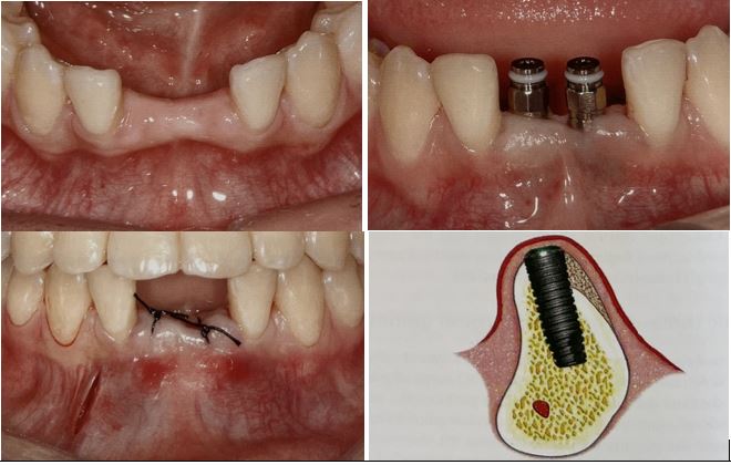
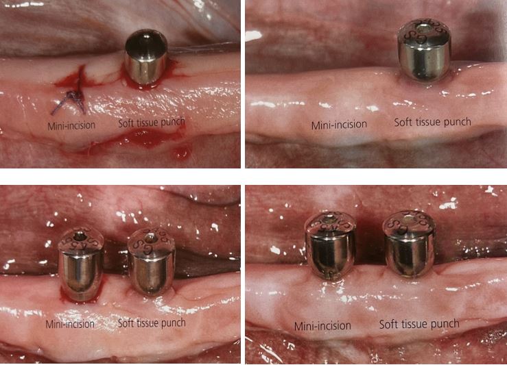
Flapless implantology Planning guided by three – dimensional images
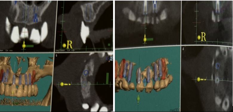
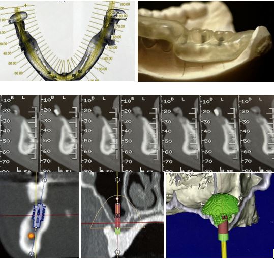
Three-dimensional planning based on CT or DVT
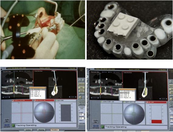
Three-dimensional planning always has to consider the requirements of the final prosthetic restoration as well as the requirements of the anatomical conditions of the implant site.
Therefore, at the beginning of the planning phase, a precise diagnostic wax-up is necessary to simulate the patient’s final restorative result concerning esthetic and function.
The diagnostic wax-up then allows fabrication of a radiographic splint.
The diagnostic wax-up of the reconstructed ridge and the restored dentition is advantageous in determining the graft width and position requirements and also for analyzing the occlusion.
Computerized tomography (CT) enables the identification of diseases and critical structures at the proposed regions, as well as the determination of bone quantity, bone quality, and the position and orientation of the dental implants.
Flapless implantology Planning guided by three – dimensional images
برنامه ریزی ایمپلنتولوژی بدون فلپ با هدایت تصاویر سه بعدی
برنامه ریزی سه بعدی بر اساس CT یا DVT
برنامه ریزی سه بعدی همیشه باید الزامات ترمیم نهایی پروتز و همچنین شرایط آناتومیکی محل ایمپلنت را در نظر بگیرد.
بنابراین، در ابتدای مرحله برنامه ریزی، یک اپیلاسیون تشخیصی دقیق برای شبیه سازی نتیجه نهایی ترمیمی بیمار در مورد زیبایی و عملکرد ضروری است.
سپس واکس آپ تشخیصی امکان ساخت اسپلینت رادیوگرافی را فراهم می کند.
واکسآپ تشخیصی برجستگی بازسازیشده و دندانهای ترمیمشده در تعیین عرض و موقعیت مورد نیاز پیوند و همچنین برای آنالیز اکلوژن مفید است.
توموگرافی کامپیوتری (CT) شناسایی بیماری ها و ساختارهای حیاتی در نواحی پیشنهادی و همچنین تعیین کمیت استخوان، کیفیت استخوان و موقعیت و جهت ایمپلنت های دندانی را امکان پذیر می سازد.

